OE-MRI for PAtients with Lung cancer (OPAL study)
Oxygen-enhanced magnetic resonance imaging (OE-MRI) for patients with lung cancer receiving chemoradiation
Project Leads: Professor James O’Connor (ICR & University of Manchester) and Professor Geoff Parker (UCL)
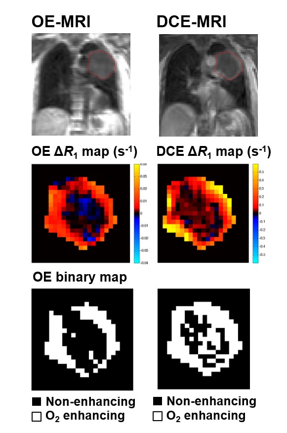
Tumour hypoxia is associated with poor survival in non-small cell lung cancer patients (NSCLC). Therefore, there is an urgent unmet need to develop non-invasive biomarkers that can detect and track changes in tumour hypoxia. Imaging biomarkers are attractive as candidates for the assessment of tumour hypoxia due to limited tissue material in radiotherapy-treated NSCLC patients.
Recently, a clinical first-in-human study, led by Professor James O’Connor at University of Manchester, demonstrated that oxygen-enhanced MRI (OE-MRI) biomarkers can be used to detect tumour hypoxia and monitor changes in hypoxia in response to treatment. 1
The NCITA Exemplar 5 OPAL study will determine the feasability of OE-MRI across different platforms and field strengths as well as multicentre repeatability of OE-MRI biomarkers. Recently, the group have reported the feasibility, repeatability and reproducibility of a dynamic T2*-sensitised OE-MRI protocol for dynamic OE-MRI of the lung in healthy volunteers.
2
The role of OE-MRI in adaptive radiotherapy planning in non-small cell lung cancer patients will also be determined. These studies are centred at The University of Manchester and University College London, before being opened up to other sites.